Try us for free
Microscopy image of a large cell prepared for segmentation

VOLKCELL

Experimental insights, without the complexity.
From raw image to actionable data in seconds, at any scale.

Products

Image analysis software

CellOpsis Live

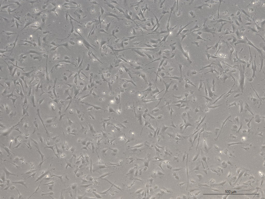
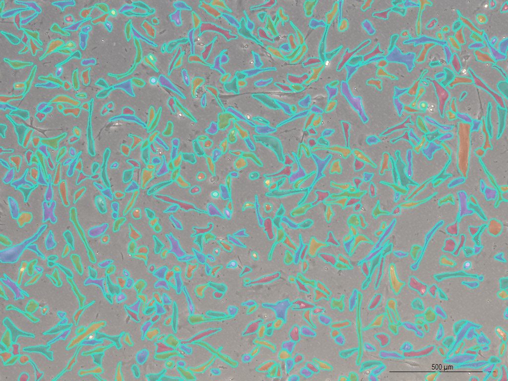
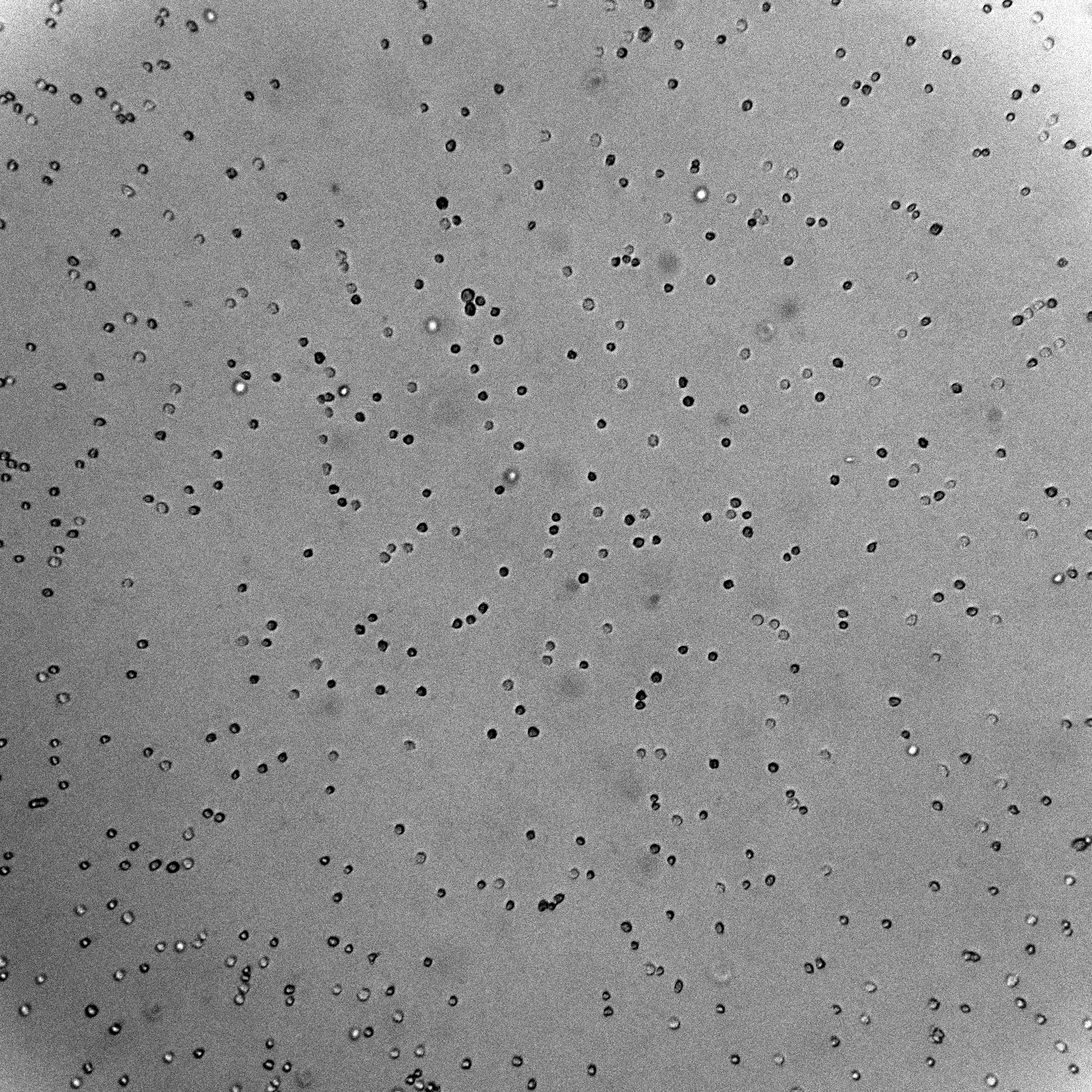
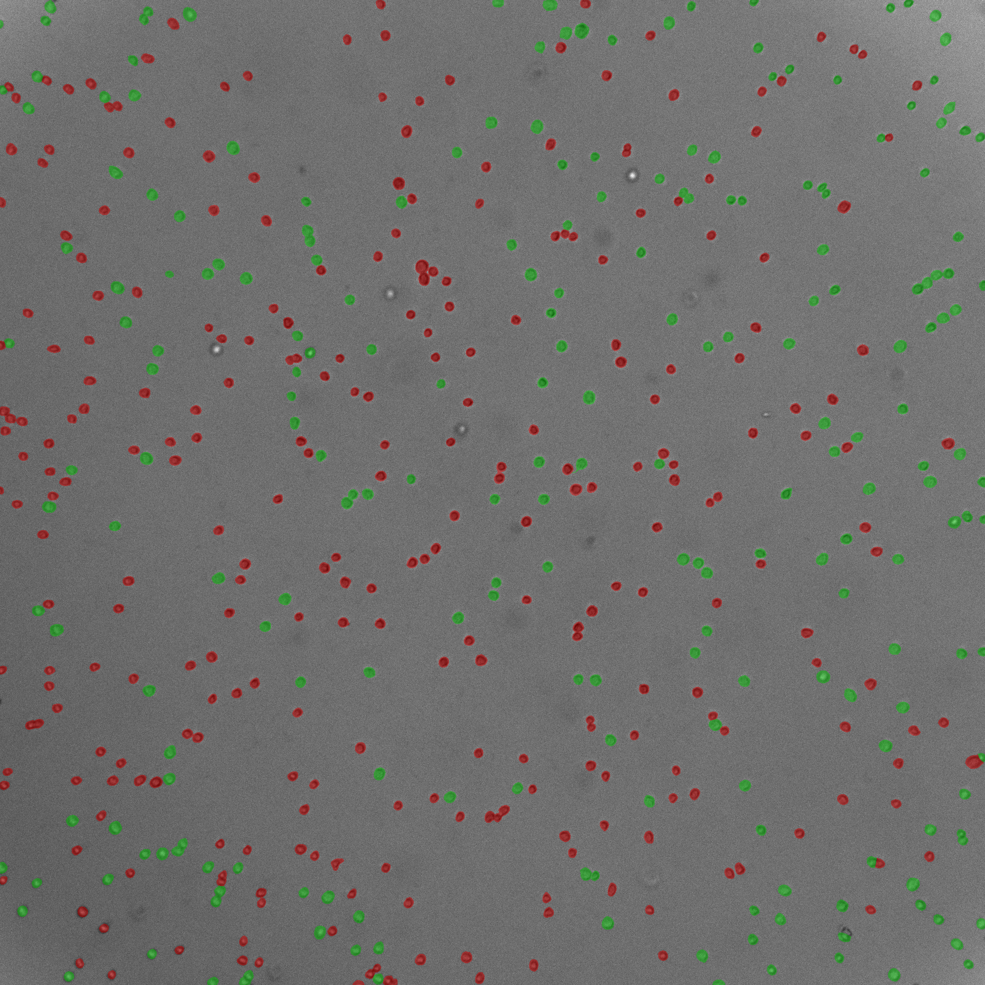
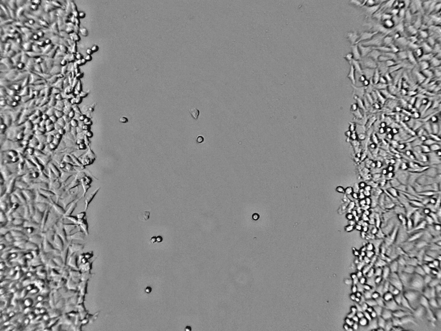
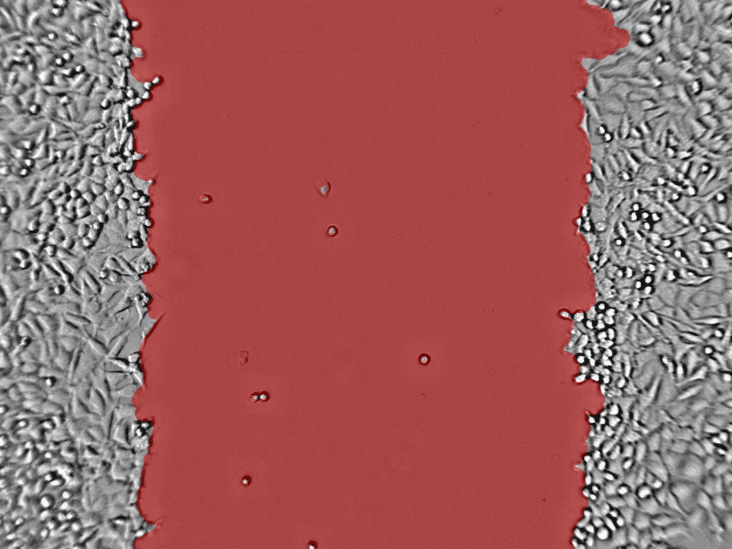
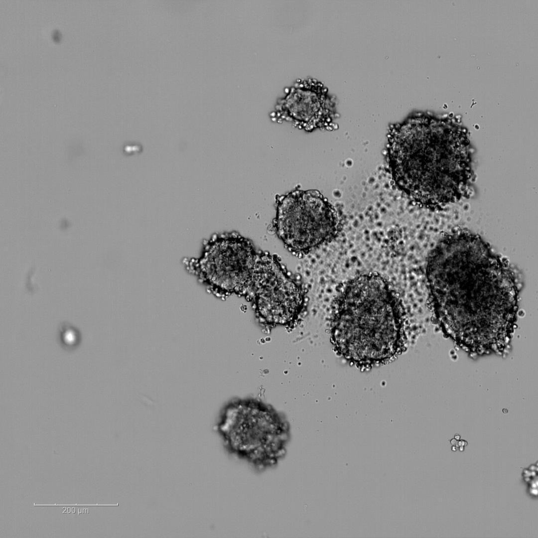
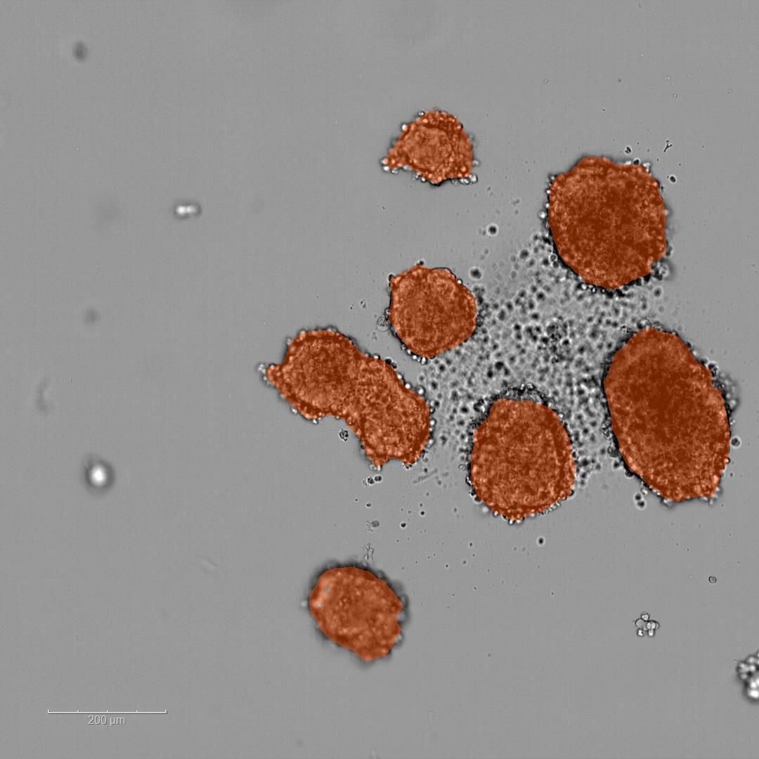
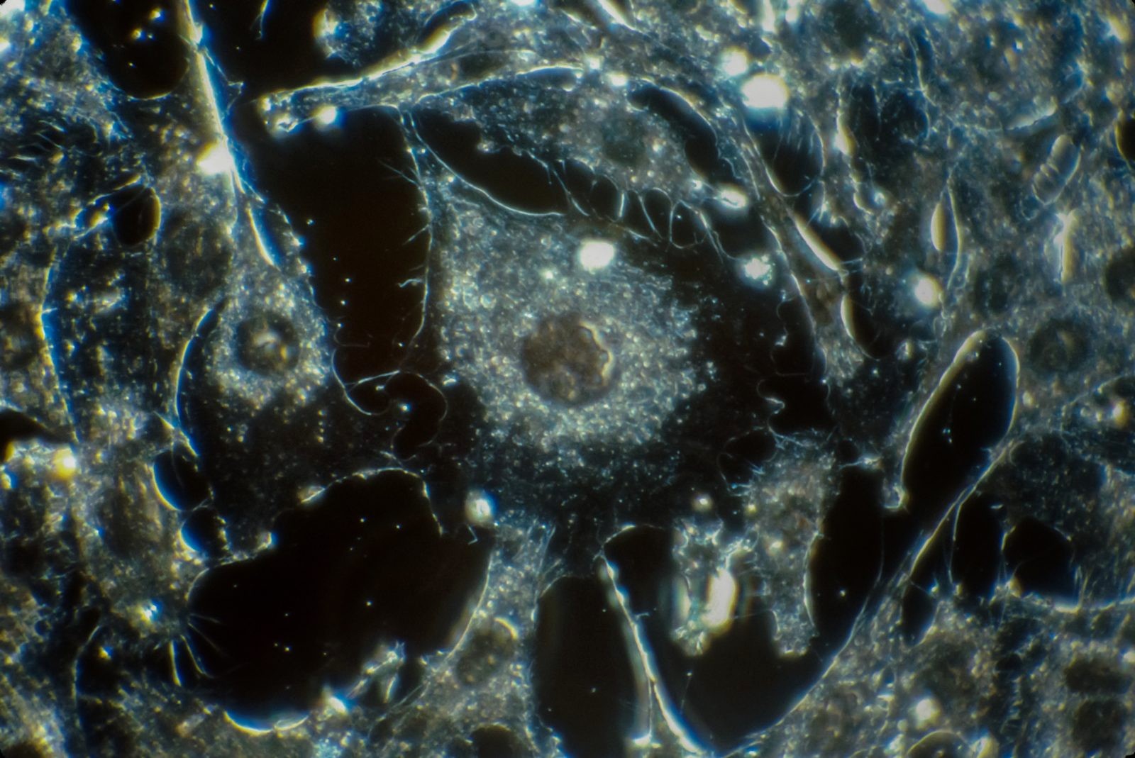
CellOpsis Live turns microscopy images into insights through a few clicks and our proprietary computer vision models.

Segment your images
Collect data
Train your own models
Track your conditions over time

Use cases for CellOpsis Live

Cell culture

Adherent cells

Track confluency, growth, and repeatable cell counts over time from label-free images.

Raw adherent cell microscopy image Adherent cell image with segmentation outlines Raw Segmented

Suspension

Suspension cells

Detect suspended cells in dense brightfield images and determine their viability over time.

Raw suspension cell microscopy image Suspension cell image with segmentation overlay Raw Segmented

Migration

Scratch assay

Follow wound closure, migration, and treatment response across timepoints.

Raw scratch assay image Scratch assay image with segmentation overlay Raw Segmented

3D culture

Spheroids

Track spheroid growth, structure, and response over time in organoid and aggregate studies.

Raw spheroid microscopy image Spheroid image with segmentation overlay Raw Segmented

Automation-ready plate reader

CellOpsis Bench

CellOpsis Bench integrates into your automation workflow with minimal space utilisation and comes ready with full automation support.

CellOpsis Bench compact plate reader
CellOpsis Bench